back
to the home page
Other
Samples
|
|
|
|
Here
are a few images of living samples taken with the AMi system.
|
|
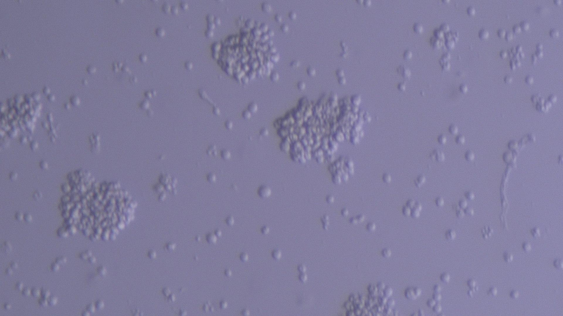
Jurkat cells (immortalized T lymphocytes) as viewed using the AMi system with the picamera and $70 zoom optics. Images acquired through the picamera can be written directly to an external computer disk and the software controls the file names and the location on the disk where the images appear.
|
The images below were taken using an earlier version of the AMi system, which incorporated an Amscope camera and a Thor Labs 16x zoom lens. This is significantly more expensive, and while the camera is triggered by the system, users have no control over the names of the individual images.
|
|
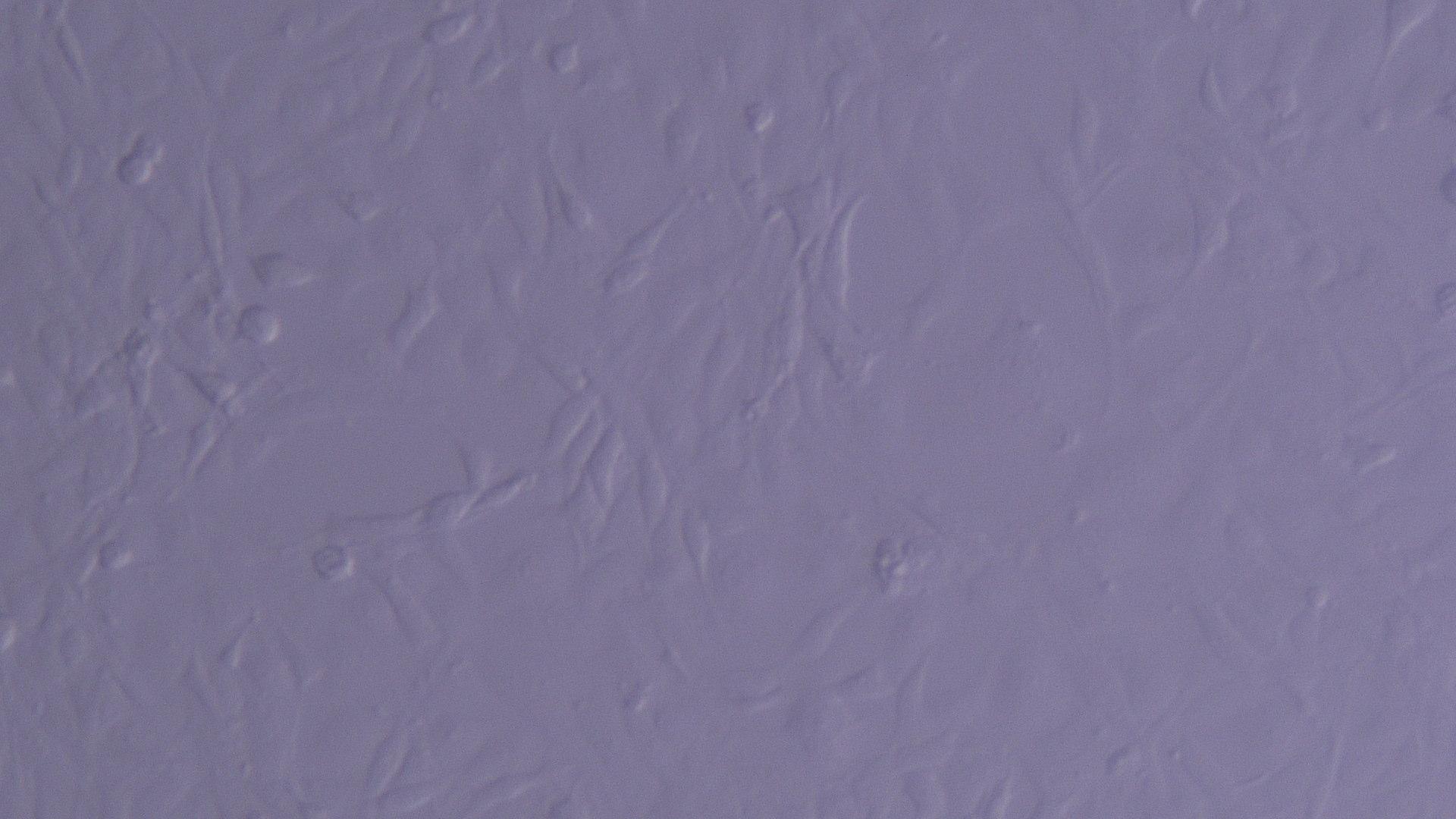
adherent fibroblast cells (7x magnification) |
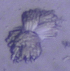
Jukat cells in suspension (5x magnification) |
|
|
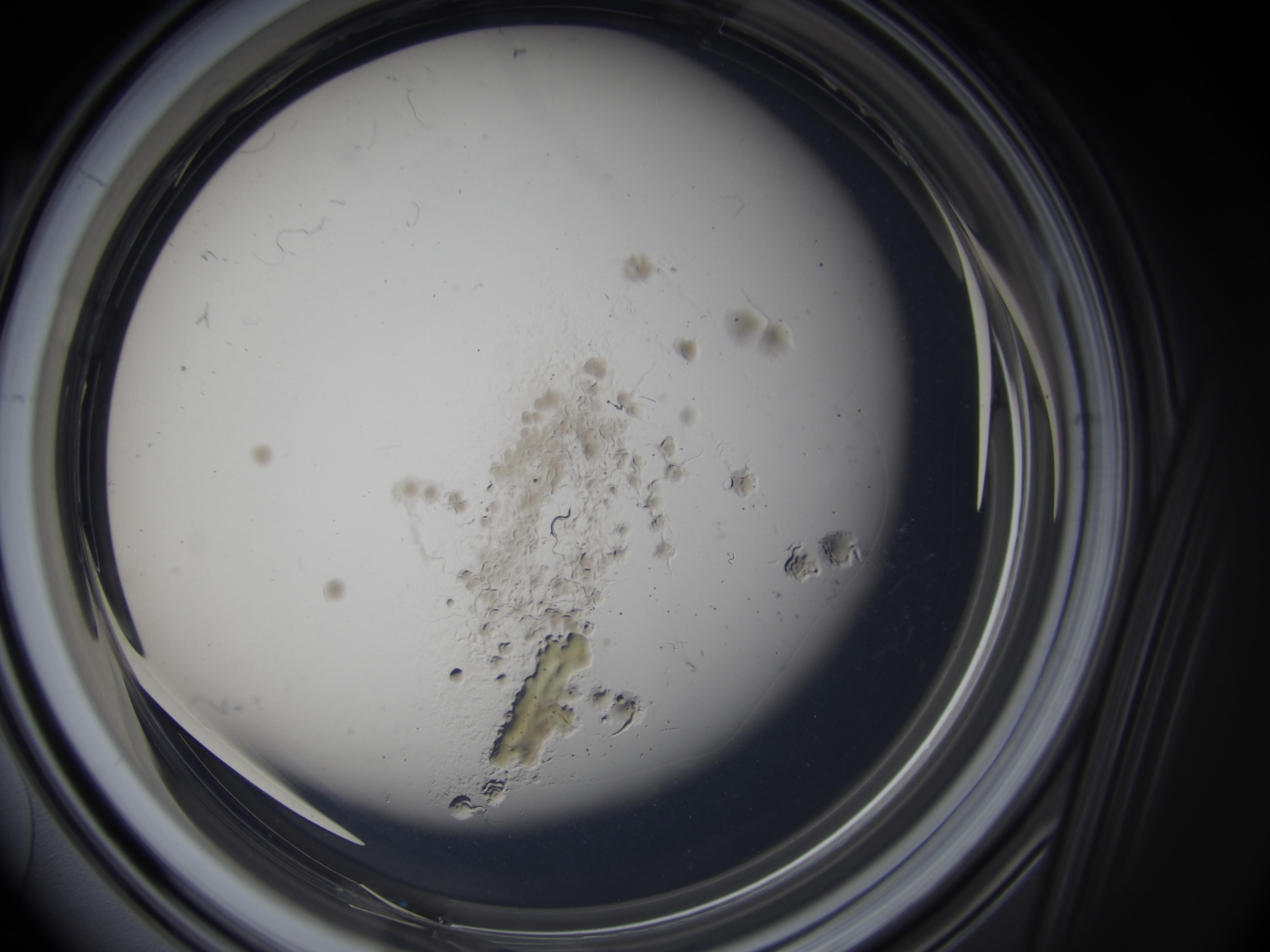
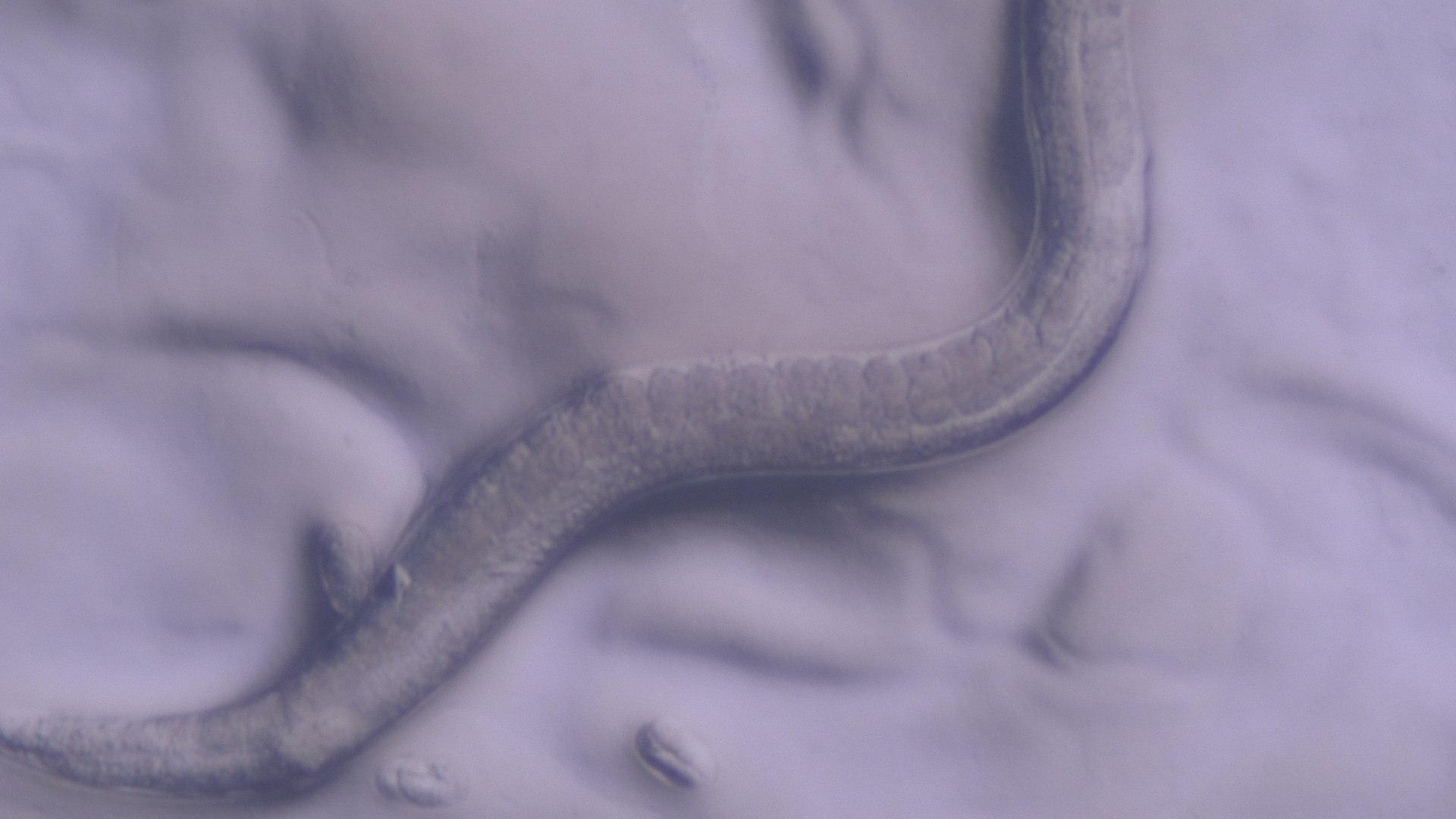
C. elegans worm at 7x magnification (note the eggs near the bottom) |
|
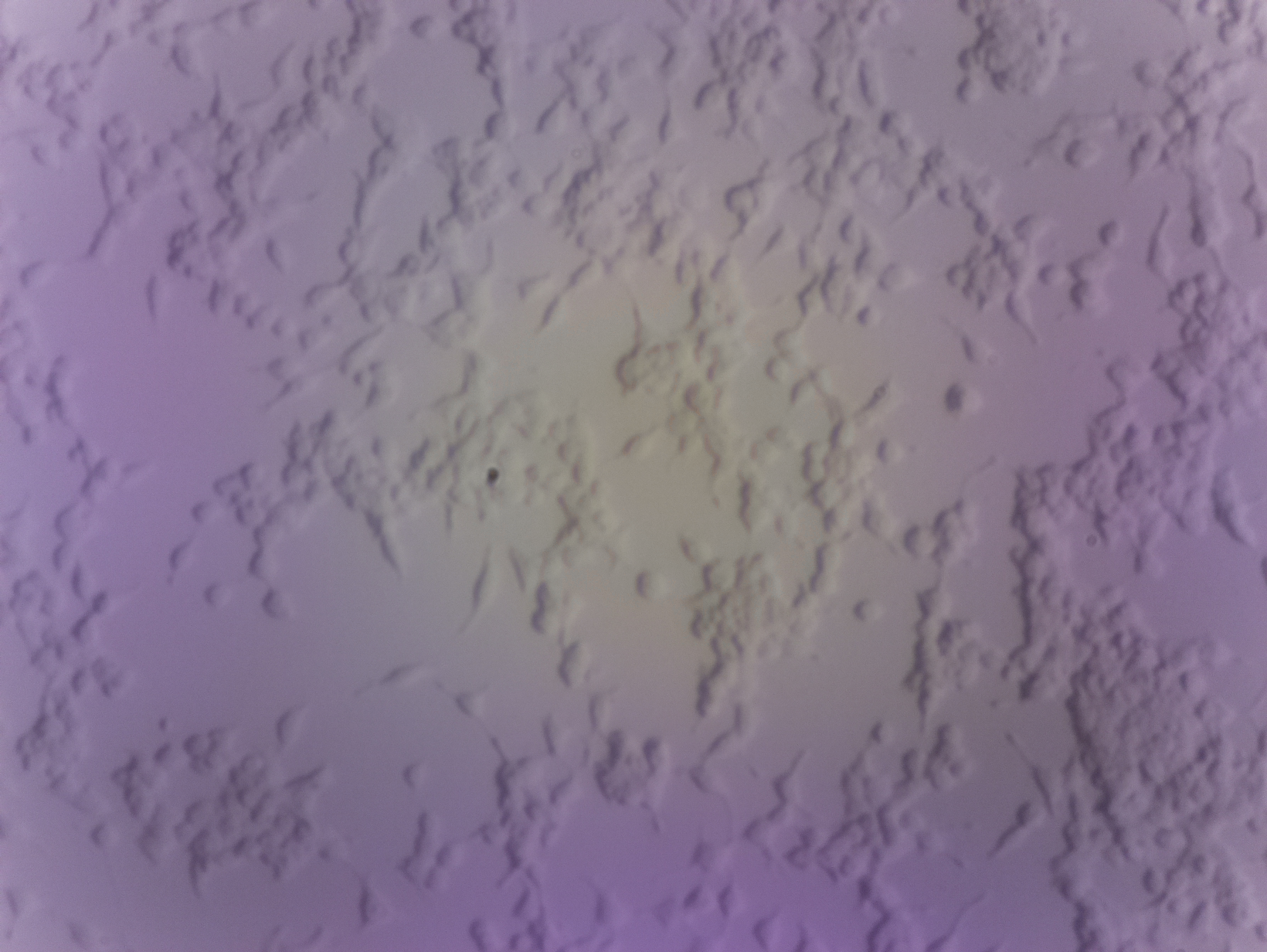
The final image (below) is of the same worm imaged above, but
using a micro 4/3 camera at 0.58x zoom. |
|